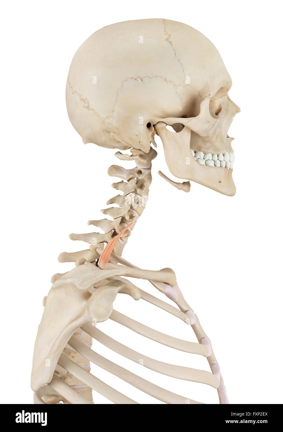
The facial skeleton is formed by the mandible, maxillae (r,l), zygomatics (r,l), and the bones that give shape to the nasal cavity: lacrimals (r,l), nasals (r,l), vomer, palatines (r,l), and the nasal conchae (r,l). The 14 bones of the facial skeleton form the entrances to the respiratory and digestive tracts. The facial skeleton, as its name suggests, makes up the face of the skull. The cranial bones compose the top and back of the skull and enclose the brain. The skull consists of the cranial bones and the facial skeleton. Skull Bones Protect the Brain and Form an Entrance to the Body. The bones of the appendicular skeleton (the limbs and girdles) “append” to the axial skeleton. The axial skeleton includes the bones that form the skull, laryngeal skeleton, vertebral column, and thoracic cage. Let’s work our way down this axis to learn about these structures and the bones that form them. The axial skeleton includes all the bones along the body’s long axis.

The appendicular skeleton includes all the bones that form the upper and lower limbs, and the shoulder and pelvic girdles. The cranial nerves in the anterior triangle are the facial, glossopharyngeal, vagus, accessory, and hypoglossal nerves.The bones of the human skeleton are divided into two groups. Some pass straight through, and others give rise to branches which innervate some of the other structures within the triangle. Numerous cranial nerves are located in the anterior triangle. The internal jugular vein can also be found within this area - it is responsible for venous drainage of the head and neck. The common carotid artery bifurcates within the triangle into the external and internal carotid branches. There are several important vascular structures within the anterior triangle. Fig 1.0 - Borders of the anterior triangle of the neck. The floor of the submental triangle is formed by the mylohyoid muscle, which runs from the mandible to the hyoid bone.


The internal jugular vein can also be found within this area – it is responsible for venous drainage of the head and neck.

The suprahyoid muscles are located superiorly to the hyoid bone, and infrahyoids inferiorly. The muscles in this part of the neck are divided as to where they lie in relation to the hyoid bone. The contents of the anterior triangle include muscles, nerves, arteries, veins and lymph nodes.


 0 kommentar(er)
